We are sampling within a phytoplankton bloom, or possibly after the peak of the bloom. When we check the water under the microscope, we can see the many different phytoplankton cells that together form a bloom.
Life in a drop of water:
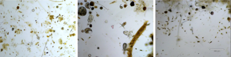
And in a higher magnification:

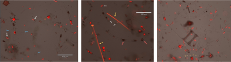
Overlay images:
There are many pennate diatoms (white arrows), and some empty frustules (dead diatoms) blue arrows.
Yellow indicates phycoerythrin autofluorescence, this pigment characterize some cyanobacteria as Trichodesmium (yellow arrows, middle image), and Richelia, both are nitrogen fixers that can be the driving force behind this bloom.
Apart from appreciating the beauty and variety of these phytoplankton, I try to isolate some of the diatoms.
It can take weeks to achieve pure cultures, but I made some progress, and started to isolate the diatoms bellow, possibly Hemiaulus, that already looks like monoculture.

All the images are from a 22N, 158W, surface seawater.